
PhD, MD, PU-PH,
alain.luciani@aphp.fr
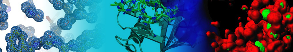
Imaging is essential in the management of cancer, especially liver cancer. The optimization of imaging, mainly by the use of cross-sectional imaging techniques, allows both morphological and functional assessment, enabling the extraction of information directly from functional and cellular tissue (cellularity, vascularization, angiogenesis, spectroscopic quantification) within tumoral lesions.
The research and development topics of the research group in translational imaging concern primarily the optimization of non-invasive imaging techniques for the preclinical detection of cancer and the study of transformation mechanisms by imaging in vivo.
Three research topics are currently being explored:
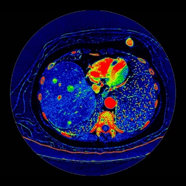
TDM imaging in spectral decomposition:
iodine / water mapping illustrating iodine enhancement in CHC nodules
Impact of hepatobiliary phase liver MRI versus Contrast-Enhanced Ultrasound after an inconclusive extracellular gadolinium-based contrast-enhanced MRI for the diagnosis of benign hepatocellular tumors
Abdom Radiol (NY). 2017
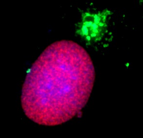
Adipose tissue macrophages: MR tracking to monitor obesity-associated inflammation
Radiology 2012
Non-invasive diagnostic imaging of hepatocellular carcinoma: targeting cellular characterization?
J Hepatol. 2011
Functional imaging of hepatocellular carcinoma using diffusion-weighted MRI and (18)F-FDG PET/CT in patients on waiting-list for liver transplantation
Cancer Imaging. 2016
Differentiation of focal nodular hyperplasia from hepatocellular adenoma: Role of the quantitative analysis of gadobenate dimeglumine-enhanced hepatobiliary phase MRI
J Magn Reson Imaging. 2015
Differentiation of focal nodular hyperplasia from hepatocellular adenomas with low-mechanical-index contrast-enhanced sonography (CEUS): effect of size on diagnostic confidence
Eur Radiol. 2015