
PhD, MD, PU-PH,
alain.luciani@aphp.fr
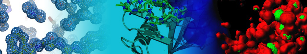
L'imagerie est incontournable dans la prise en charge des cancers, en particulier les cancers du foie. Les optimisations instrumentales en imagerie, principalement en imagerie de coupe, permettent une approche à la fois morphologique et fonctionnelle, permettant d'extraire des informations sur le fonctionnement tissulaire et cellulaire (cellularité, vascularisation, angiogenèse, quantification spectroscopique) directement au sein des lésions tumorales.
Les thématiques de recherche et développement en imagerie translationnelle au sein de l'équipe de recherche concernent avant tout l'optimisation instrumentale de techniques d'imagerie non invasive pour la détection préclinique des cancers et pour la compréhension des mécanismes de transformation par l'imagerie in vivo.
Trois axes de recherche sont actuellement portés :
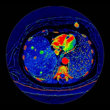
Imagerie TDM en décomposition spectrale :
cartographie iode/eau illustrant les prises de contraste iodées au sein de nodules de CHC
Impact of hepatobiliary phase liver MRI versus Contrast-Enhanced Ultrasound after an inconclusive extracellular gadolinium-based contrast-enhanced MRI for the diagnosis of benign hepatocellular tumors
Abdom Radiol (NY). 2017
Adipose tissue macrophages: MR tracking to monitor obesity-associated inflammation
Radiology 2012
Non-invasive diagnostic imaging of hepatocellular carcinoma: targeting cellular characterization?
J Hepatol. 2011
Functional imaging of hepatocellular carcinoma using diffusion-weighted MRI and (18)F-FDG PET/CT in patients on waiting-list for liver transplantation
Cancer Imaging. 2016
Differentiation of focal nodular hyperplasia from hepatocellular adenoma: Role of the quantitative analysis of gadobenate dimeglumine-enhanced hepatobiliary phase MRI
J Magn Reson Imaging. 2015
Differentiation of focal nodular hyperplasia from hepatocellular adenomas with low-mechanical-index contrast-enhanced sonography (CEUS): effect of size on diagnostic confidence
Eur Radiol. 2015